Home
/ Pelvic Anatomy Posterior View : Pelvic Anatomy Related To Shoulder Dystocia - 1.2 rectal anatomy , lateral view fig.
Pelvic Anatomy Posterior View : Pelvic Anatomy Related To Shoulder Dystocia - 1.2 rectal anatomy , lateral view fig.
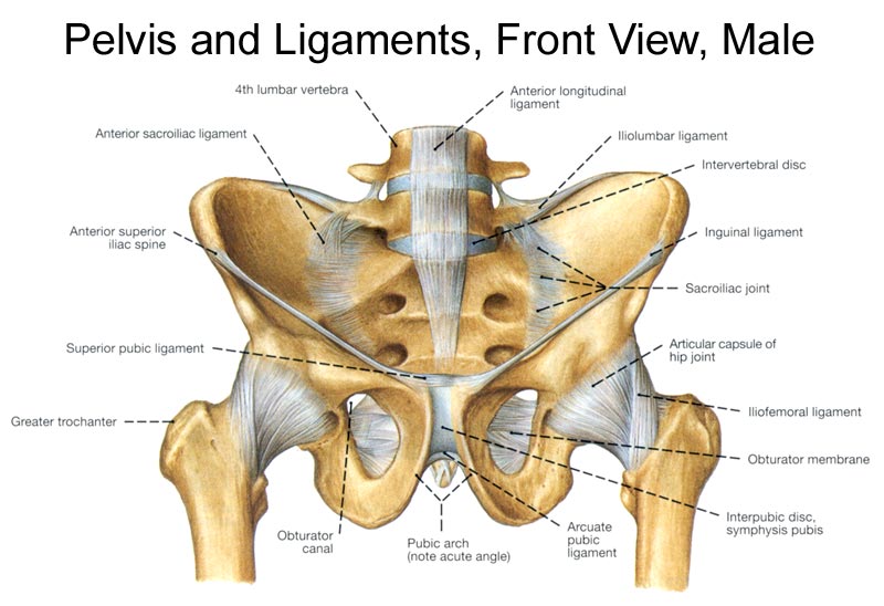
Pelvic Anatomy Posterior View : Pelvic Anatomy Related To Shoulder Dystocia - 1.2 rectal anatomy , lateral view fig.. The iliopectineal, or arcuate, line divides the pelvis into the upper pelvis (which is part of the abdomen and the lower pelvis) and the true pelvis. 16.2b ilium pubic tubercle ischial spine iliac crest iliac fossa anterior superior iliac spine (asis) greater sciatic notch lesser sciatic notch ischial tubercle ischial ramus anterior inferior iliac spine (aiis) netter's atlas of human. During pregnancy, temporary changes take place in the ligaments that permit both movement of the joints and enlargement of the pelvic cavity. The posterior pelvis transmits force from the body to the lower extremities while the pubis functions primarily as a strut. You need to get 100% to score the 8 points available.
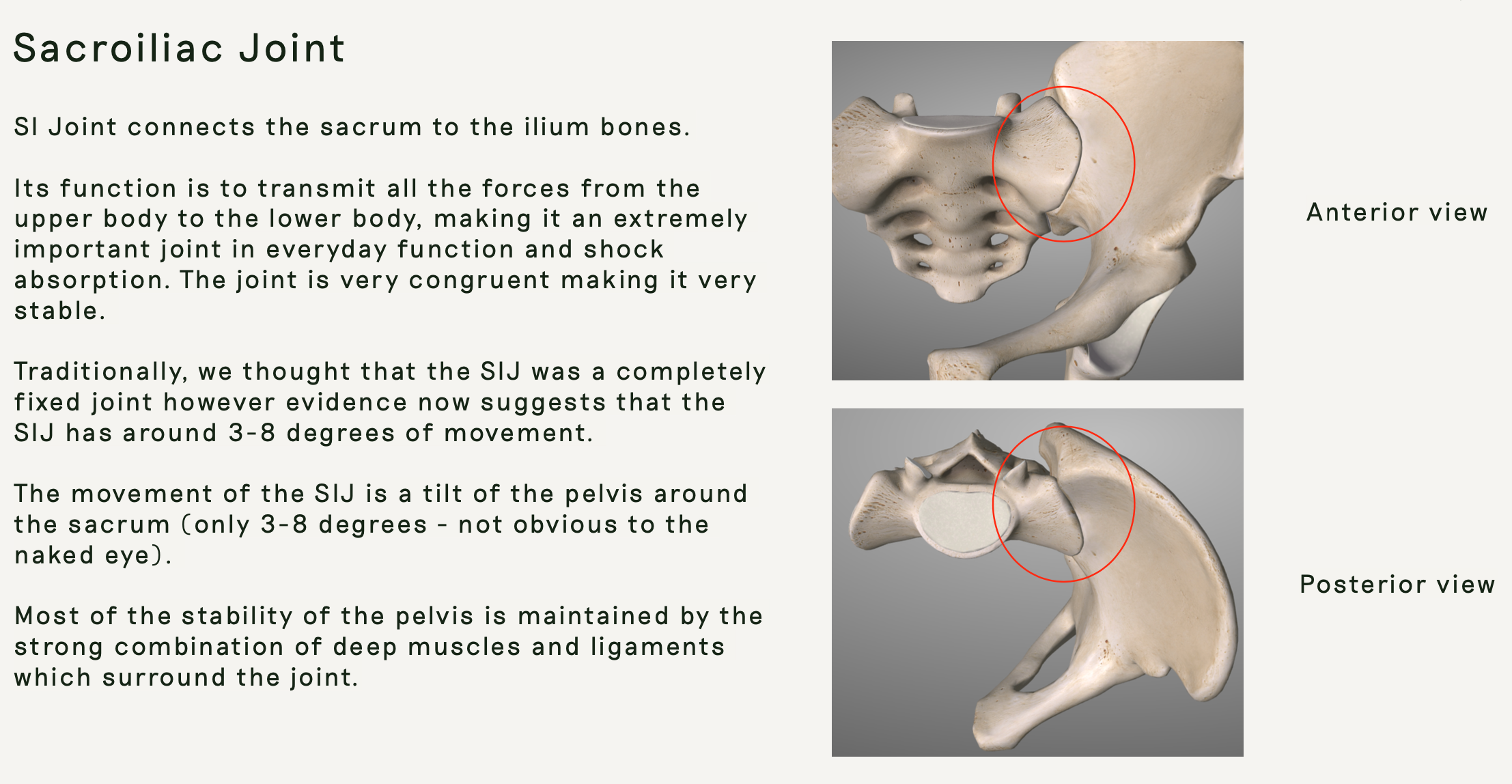
1.2 rectal anatomy , lateral view fig. Secondly, the obturator oblique view demonstrating the anterior column of the pelvis along with the posterior wall of the acetabulum. Posterior view of the lumbar spine and pelvis. The vertebral column of the lower back includes the five lumbar vertebrae, the sacrum, and the coccyx. The two pelvic bones are connected anteriorly by the pubic symphysis, while posteriorly they articulate with the pelvic spine to form the sacroiliac joints.
Pelvic Anatomy Good Day Pilates from images.squarespace-cdn.com Ebraheim's educational animated video describes the anatomy of the pelvis, the bony structures, ligaments, muscles, blood supply, and nerves.this video a. The pelvic spine is the posterior portion of the pelvis below the lumbar spine, composed of the sacrum and coccyx. This mri male pelvis axial cross sectional anatomy tool is absolutely free to use. The lumbar spine is composed of five vertebrae, named l1 to l5 from superior to inferior. The main function of the pelvic floor muscles are: The posterior ring structures are responsible for the majority of pelvic ring stability. You need to get 100% to score the 8 points available. The structure of the pelvis supports the contents of the abdomen while also helping to transfer the weight from the spine to the lower limbs.
The pelvic bones are smaller and.
Bone and ligaments of pelvis posterior view in this image, you will find the posterior superior iliac spine, iliac crest, tubercle of the iliac crest, anterior superior iliac spine, greater sciatic foramen, the acetabular margin in it. Anatomynote.com found pelvic region posterior view from plenty of anatomical pictures on the internet. We think this is the most useful anatomy picture that you need. Bones and ligaments of the female pelvis this image shows the posterior back view of the female pelvic brim (the bones and ligaments that forms the pelvic region in the female) The posterior ring structures are responsible for the majority of pelvic ring stability. Secondly, the obturator oblique view demonstrating the anterior column of the pelvis along with the posterior wall of the acetabulum. You need to get 100% to score the 8 points available. The main function of the pelvic floor muscles are: Used freely under public domain. To support the abdominal and pelvic viscera. The two pelvic bones are connected anteriorly by the pubic symphysis, while posteriorly they articulate with the pelvic spine to form the sacroiliac joints. Connects iliac and obturator systems. To maintain the continence of urine and faeces.
The symphyseal ligaments, which hold the pubis together, resist external rotation and account for only 15% of the stability to the entire ring. Transverse ligaments, like the short posterior sacroiliac and the anterior sacroiliac ligaments along with the iliolumbar and sacrospinous ligaments, resist rotational forces. Anatomynote.com found pelvic region posterior view from plenty of anatomical pictures on the internet. The superior border of the ilium. And the thigh to extend on the pelvis.
Bony Pelvis Anatomy Bone And Spine from boneandspine.com During pregnancy, temporary changes take place in the ligaments that permit both movement of the joints and enlargement of the pelvic cavity. Connects iliac and obturator systems. This becomes important during parturition. The lumbar spine is composed of five vertebrae, named l1 to l5 from superior to inferior. To maintain the continence of urine and faeces. The posterior ring structures are responsible for the majority of pelvic ring stability. Divides distal and posterior near the si joint into. Used freely under public domain.
The judet view is comprised of two projections, first the iliac oblique for assessment of the posterior column and anterior wall of the acetabulum;
We think this is the most useful anatomy picture that you need. Ebraheim's educational animated video describes the anatomy of the pelvis, the bony structures, ligaments, muscles, blood supply, and nerves.this video a. The lumbar spine is composed of five vertebrae, named l1 to l5 from superior to inferior. The pelvic bones are smaller and. Bone and ligaments of pelvis posterior view in this image, you will find the posterior superior iliac spine, iliac crest, tubercle of the iliac crest, anterior superior iliac spine, greater sciatic foramen, the acetabular margin in it. Leads to superior guteal artery and other branches. Transverse ligaments, like the short posterior sacroiliac and the anterior sacroiliac ligaments along with the iliolumbar and sacrospinous ligaments, resist rotational forces. Used freely under public domain. 1.3 rectal anatomy , ap view fig. Injury in pelvic fractures can account for majority of blood loss. Ths anatomy pelvis posterior view learn by taking a quiz; The superior border of the ilium. The bones of the pelvis and lower back work together to support the body's weight, anchor the abdominal and hip muscles, and protect the delicate vital organs of the vertebral and abdominopelvic cavities.
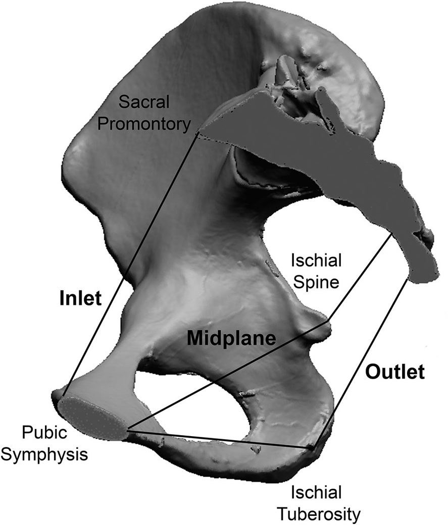
The judet view is comprised of two projections, first the iliac oblique for assessment of the posterior column and anterior wall of the acetabulum; Leads to superior guteal artery and other branches. 16.2b ilium pubic tubercle ischial spine iliac crest iliac fossa anterior superior iliac spine (asis) greater sciatic notch lesser sciatic notch ischial tubercle ischial ramus anterior inferior iliac spine (aiis) netter's atlas of human. The pelvic region is the area between the trunk — or main body — and the lower extremities, or legs. The lumbar spine is composed of five vertebrae, named l1 to l5 from superior to inferior.
Pelvis Anatomy Chapter 1 The Evolutionary Biology Of The Human Pelvis from static.cambridge.org This mri male pelvis axial cross sectional anatomy tool is absolutely free to use. Ths anatomy pelvis posterior view learn by taking a quiz; Terms in this set (14) iliac crest. Connects iliac and obturator systems. 1.2 rectal anatomy , lateral view fig. The pelvis consists of the sacrum, the coccyx, the ischium, the ilium, and the pubis. The pelvis is composed of the two pelvic bones and the sacrum and coccyx. Together, they form the part of the pelvis called the pelvic girdle.
You need to get 100% to score the 8 points available.
Ebraheim's educational animated video describes the anatomy of the pelvis, the bony structures, ligaments, muscles, blood supply, and nerves.this video a. The pelvis consists of the sacrum, the coccyx, the ischium, the ilium, and the pubis. The superior border of the ilium. Bones and ligaments of the female pelvis this image shows the posterior back view of the female pelvic brim (the bones and ligaments that forms the pelvic region in the female) The posterior pelvis transmits force from the body to the lower extremities while the pubis functions primarily as a strut. Injury in pelvic fractures can account for majority of blood loss. There are two hip bones, one on the left side of the body and the other on the right. During pregnancy, temporary changes take place in the ligaments that permit both movement of the joints and enlargement of the pelvic cavity. Secondly, the obturator oblique view demonstrating the anterior column of the pelvis along with the posterior wall of the acetabulum. The pelvis is composed of the two pelvic bones and the sacrum and coccyx. Use the mouse scroll wheel to move the images up and down alternatively use the tiny arrows (>>) on both side of the image to move the images.>>) on both side of the image to move the images. Major ligaments and notches of the female pelvis, posterior view. The posterior ring structures are responsible for the majority of pelvic ring stability.
Injury in pelvic fractures can account for majority of blood loss pelvic anatomy. During pregnancy, temporary changes take place in the ligaments that permit both movement of the joints and enlargement of the pelvic cavity.